Key takeaways:
The most common sign of skin cancer is a new or changing spot on your skin that doesn’t go away on its own.
Skin cancer usually appears on sun-exposed skin, but that’s not always the case. So it’s a good habit to regularly check all of your skin.
If you’re worried about a spot on your skin, trust your gut and visit your dermatologist or your primary care provider to get it checked out.
Getting new bumps on your skin is common. Most of the time, it’s just something like a pimple or cyst and is nothing to worry about. But how do you know when a change on your skin is serious? In other words, when should you worry about skin cancer and take action?
These are great questions to ask. After all, skin cancer is the most common type of cancer in the U.S. and affects people of all skin tones. Fortunately, skin cancer is very curable when you catch it early. And there are steps you can take to spot skin cancer early — including checking your skin regularly and knowing what clues to look for.
What does early skin cancer look like?
Precancerous spots are called actinic keratosis. They’re common on sun-damaged skin. A small percentage of these will develop into a type of skin cancer called squamous cell carcinoma (more on this below).
Early skin cancer can be hard to spot. It may look like a dry patch of skin, or an acne-like spot that doesn’t go away. Here are some pictures to help you spot early-stage skin cancer.
Basal cell carcinoma (BCC)
Basal cell carcinoma (BCC) is the most common type of skin cancer. It usually occurs on sun-exposed skin, like the face, ears, head, and neck. There are a few different types of BCC.
Here are the most common ones, with pictures and descriptions to help you spot them.
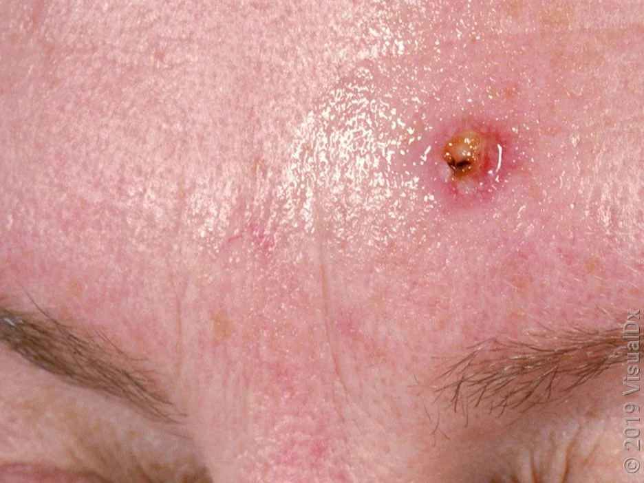
Nodular basal cell carcinoma
This is the most common type of BCC. It forms a smooth, pink or brown skin bump with raised edges. There may be a sore in the middle that doesn’t heal.

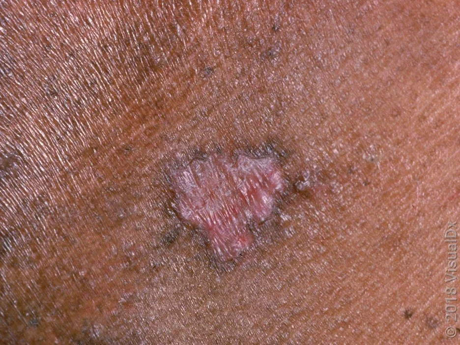
Morpheaform basal cell carcinoma
This type forms a flat scar-like area that’s shiny and smooth. They can feel firm to the touch.

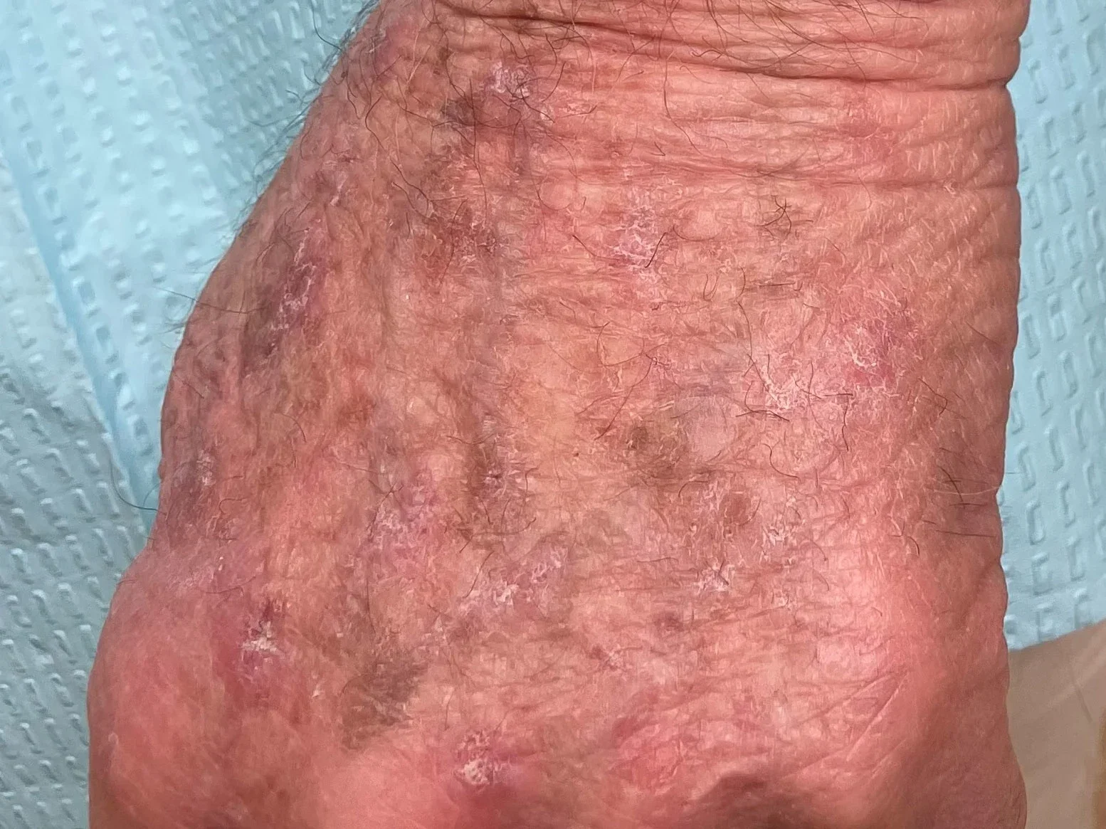
Superficial basal cell carcinoma
This type forms a rough, reddish or brown, slightly raised patch that doesn’t go away. It’s common on the torso, arms, and legs, and it can spread wide, but doesn’t go deep into the skin.

Pigmented basal cell carcinoma
This type of BCC is partly or entirely brown in color (pigmented). In people with darker skin tones, this type accounts for about half of all BCCs.

Images of squamous cell carcinoma (SCC)
Squamous cell carcinoma (SCC) is the second most common type of skin cancer. It can occur anywhere on the body. But this type of skin cancer usually happens on sun-exposed skin. This includes areas like the face, ears, neck, and back of hands. SCC can also develop in scars or chronically inflamed skin.
What does squamous cell carcinoma look like?
Here are some signs of SCC to look for:
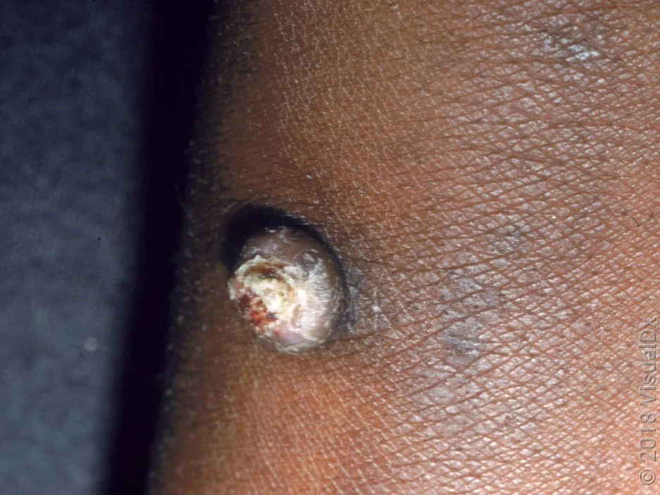
Wart-like growth that’s crusty
Sore that doesn’t heal and can bleed
Crater-like growth with a central depression
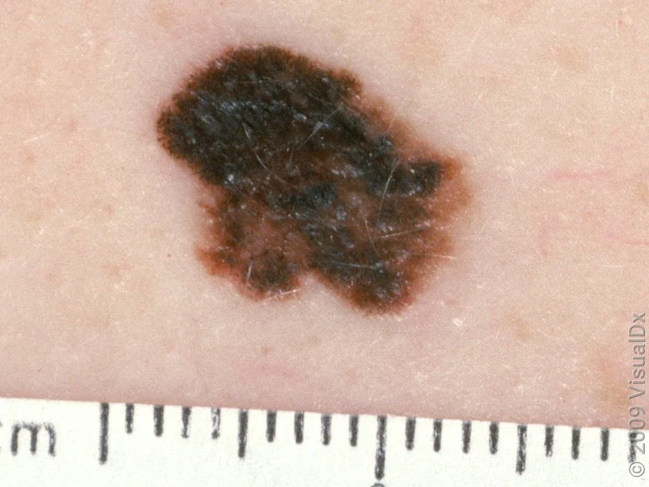
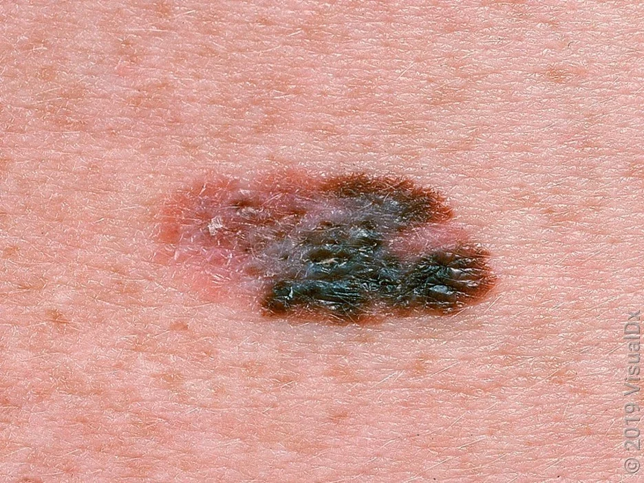
Melanoma
Melanoma is the least common but most serious type of skin cancer. It’s the most serious because it’s more likely to spread (metastasize) to other organs in your body. And that makes it much more difficult to treat. About 20% to 30% of melanomas develop in a mole you already have, so it’s important to know what changes to look for.
What does melanoma look like?
A melanoma can look like a brown or black spot. But it can also look blue, pink, or the same color as your skin. The spot can be flat, or it may become bumpy and raised. Melanoma can develop anywhere on your skin — including areas that aren’t exposed to the sun.
The “ABCDE rule” and “ugly duckling sign” can help you you spot early signs of melanoma:
Asymmetry: A mole where one half doesn’t look like the other half
Border: A mole that has a bumpy or irregular border
Color: A spot that has different colors (like tan, black, white, or pink)
Diameter: A mole that’s larger than a pencil eraser
Evolving: Any change in an existing mole or skin lesion
Ugly duckling: Any mole that doesn’t look like your other moles
There are four main types of melanoma. Here’s what these different types look like, and the parts of the body where they’re more likely to appear.
Superficial spreading melanoma
This type of melanoma can appear anywhere, but it’s more common on legs in women and on the trunk in men. It’s also common on the upper back in both men and women.

Nodular melanoma
This type often develops on the trunk, arms, or legs. It can also appear on the scalp in older men.

Lentigo maligna melanoma
This type of melanoma usually develops in older people on sun-damaged skin, like the face, ears, and back. It can grow for years in the upper skin level before becoming invasive (growing deeper into the skin).

Acral lentiginous melanoma
This is the most common type of melanoma in people with darker skin tones and people with Asian ancestry. But it can happen in any skin type. It’s common on the palms, soles of the feet, or under the nail (subungual).

Skin conditions that you may mistake for skin cancer
Not all skin changes mean cancer. Some common skin conditions can mimic skin cancer. And sometimes the only way to tell them apart is with a skin biopsy. If you’re ever in doubt, it’s best to get checked out. Here are some examples:
Sebaceous hyperplasia
When should you see a healthcare professional about a worrisome mole or growth?
If you notice a change in your skin that’s not normal for you, it’s best to have a dermatologist or other healthcare professional check it out. This is especially true if you have a spot that you think could be melanoma.
Keep in mind that most skin spots that aren’t cancer — like a pimple or cut — will go away on their own within a couple of weeks. Skin cancer never goes away on its own. And the earlier you catch it, the better.
How do you diagnose skin cancer?
The diagnosis for skin cancer usually includes a skin check and then a skin biopsy — a minor, in-office surgical procedure. They’ll use a small needle to numb your skin and then remove the spot. You may get a stitch to help the area heal. Your healthcare professional will send the tissue to a lab, where a pathologist evaluates it under the microscope.
Skin biopsies are usually done by a dermatologist, but your primary healthcare professional may also do it. If it’s skin cancer, you’ll need another surgery to remove the spot.
The bottom line
Skin cancer usually shows up as a visible change in your skin — but it’s not always a mole. The most common types of skin cancer don’t look like moles. They might be a new or changing spot that doesn’t heal, a crusty lesion, a patch, or a wart-like growth.
Skin cancer is much easier to treat when you catch it early. This means it’s important to check all your skin regularly and to be familiar with signs of possible skin cancer. Skin cancer is easy to diagnose. If you’re worried about a spot on your skin, it’s best to trust your gut and get it checked out.

Why trust our experts?


Images used with permission from VisualDx (www.visualdx.com).
References
AIM at Melanoma Foundation. (n.d.). Acral lentiginous melanoma.
American Academy of Dermatology Association. (n.d.). Actinic keratosis: Overview.
American Academy of Dermatology Association. (n.d.). Birthmarks: Overview.
American Academy of Dermatology Association. (n.d.). Seborrheic keratoses: Overview.
American Academy of Dermatology Association. (n.d.). What to look for: ABCDEs of melanoma.
American Academy of Dermatology Association. (2021). What can get rid of age spots?
American Academy of Dermatology Association. (2023). Find skin cancer: How to perform a skin self-exam.
American Academy of Dermatology Association. (2025). Skin cancer.
American Academy of Dermatology Association. (2025). Warts: Diagnosis and treatment.
Ludmann, P. (2022). What is a skin biopsy? American Academy of Dermatology Association.
McDaniel, B., et al. (2024). Basal cell carcinoma. StatPearls.
Skin Cancer Foundation. (2025). Basal cell carcinoma overview: The most common skin cancer.
Skin Cancer Foundation. (2025). Basal cell carcinoma warning signs & pictures: What does basal cell carcinoma look like?
Skin Cancer Foundation. (2025). Melanoma overview: A dangerous skin cancer.
Skin Cancer Foundation. (2025). Melanoma warning signs: What you need to know about early signs of skin cancer.
Skin Cancer Foundation. (2025). Squamous cell carcinoma warning signs & pictures: What does squamous cell carcinoma look like?
Watson, M., et al. (2011). Melanoma surveillance in the United States: Overview of methods. Journal of the American Academy of Dermatology.